
We perform EKG/ECG (electrocardiogram), ABI (ankle-brachial index), ultrasound, mammogram, venous doppler, vein mapping, and bone density testing for osteoporosis.

-
EKG/ECG (electrocardiogram)
Records the electrical signals in your heart. It's a common test used to detect heart problems and monitor the heart's status in many situations. An EKG/ECG is a noninvasive, painless test with quick results. During an EKG/ECG, sensors (electrodes) that can detect the electrical activity of your heart are attached to your chest and sometimes your limbs. These sensors are usually left on for just a few minutes.

-
ABI (ankle-brachial index)
Test is a quick, noninvasive way to check your risk of peripheral artery disease (PAD). Peripheral artery disease is a condition in which the arteries in your legs or arms are narrowed or blocked. People with the peripheral artery disease are at an increased risk of heart attack, stroke, poor circulation and leg pain. The ankle-brachial index test compares your blood pressure measured at your ankle with your blood pressure measured at your arm. A low ankle-brachial index number can indicate narrowing or blockage of the arteries in your legs, increasing your risk of circulatory problems, and possibly causing heart disease or stroke. The ankle-brachial index test is sometimes recommended as part of a series of three tests, including the carotid ultrasound and abdominal ultrasound, to check for blocked or diseased arteries.

-
Ultrasound
Is an imaging method that uses high-frequency sound waves to produce images of structures within your body. The images can provide valuable information for diagnosing and treating a variety of diseases and conditions. Most ultrasound examinations are done using an ultrasound device outside your body, though some involve placing a device inside your body. Additional imaging, such as CT, MRI, or mammography, may be recommended.

-
Mammogram
Is an X-ray image of your breasts used to screen for breast cancer. Mammograms play a key role in early breast cancer detection and help decrease breast cancer deaths.During a mammogram, your breasts are compressed between two firm surfaces to spread out the breast tissue. Then an X-ray captures black-and-white images of your breasts that are displayed on a computer screen and examined by a doctor who looks for signs of cancer. A mammogram can be used either for screening or for diagnostic purposes. How often you should have a mammogram depends on your age and your risk of breast cancer.
-
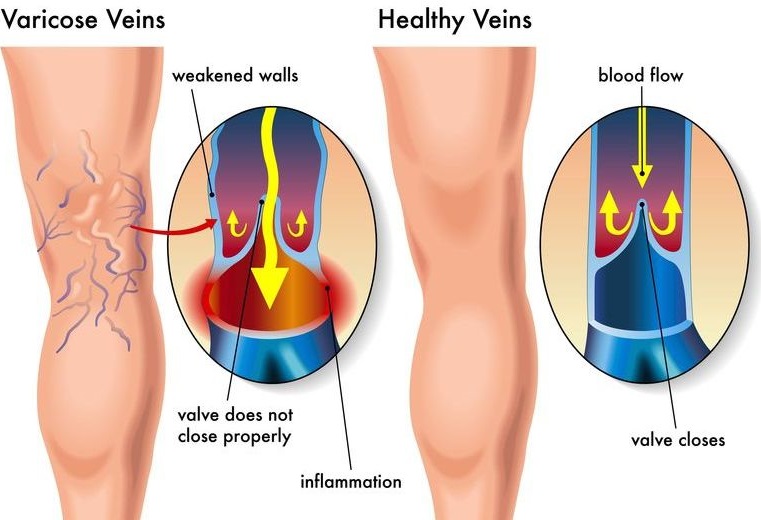
Venous Doppler
Is a special ultrasound technique that evaluates blood as it flows through a blood vessel, including the body's major arteries and veins in the abdomen, arms, legs, and neck.
-
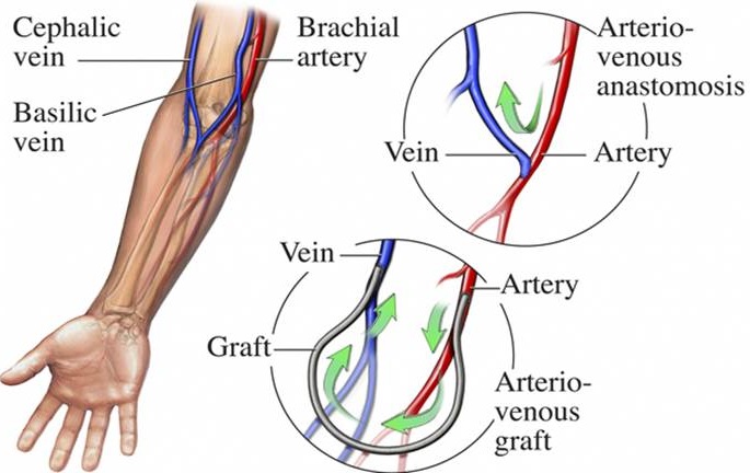
Vein Mapping
Is a test which utilizes high-frequency sound waves to obtain images of veins which are being evaluated for surgical removal or alteration. This test is used to determine if the veins are suitable for the desired surgical procedure. The technologist applies an acoustic gel on your arm or leg over the area to be viewed. Then a painless instrument called a transducer is gently moved across that portion of your body to visualize the inside of the vessels. The technician may mark on your arms to identify the location of your veins. These marks must not be removed until your doctor has directed you to do so.

-
Bone Density test
Determines if you have osteoporosis, a disorder characterized by bones that are more fragile and more likely to break. In the past, osteoporosis would be suspected only after you broke a bone. By that time, however, your bones could be quite weak. A bone density test enhances the accuracy of calculating your risk of breaking bones. A bone density test uses X-rays to measure how many grams of calcium and other bone minerals are packed into a segment of bone. The bones that are most commonly tested are in the spine, hip and sometimes the forearm.
Email Newsletters:


- Email: info@saludyvidapa.com